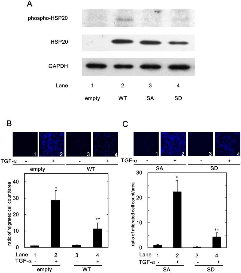
Fig 1. Phosphorylated HSP20 levels in the wild-type- and the mutants-HSP20 transfected HuH7 cells, and suppression of the TGF-α-induced HuH7 cell migration by HSP20.
(A), The protein expression of the wild-type- and the mutants-HSP20 in the HuH7 cells were determined by Western blotting using HSP20 antibodies and phospho-HSP20 (serine 16) antibodies. HuH7 cells were stably transfected either with control empty-, wild-type-HSP20-expressing (WT), its alanine mutant-expressing (SA) or its aspartate mutant-expressing (SD) vectors. The empty vector- or the WT-HSP20 transfected HuH7 cells (B), or the SA- or the SD-HSP20 overexpressed HuH7 cells (C) were stimulated by 3 ng/ml of TGF-α or vehicle for 24 h. The migrated cells were fixed with paraformaldehyde and stained with DAPI for the nucleus (blue signal). The cells were photographed by fluorescent microscopy at a magnification of 20× (upper panel). The migrated cell numbers were counted and plotted as the ratio to the mean of migrated numbers of the control empty vector-transfected cells (B), or the SA-HSP20 overexpressed cells (C) without TGF-α stimulation (lower panel). Each value represents the means ± SD (n = 3). *P<0.05, compared to the value of lane 1. **P<0.05, compared to the value of lane 2.

