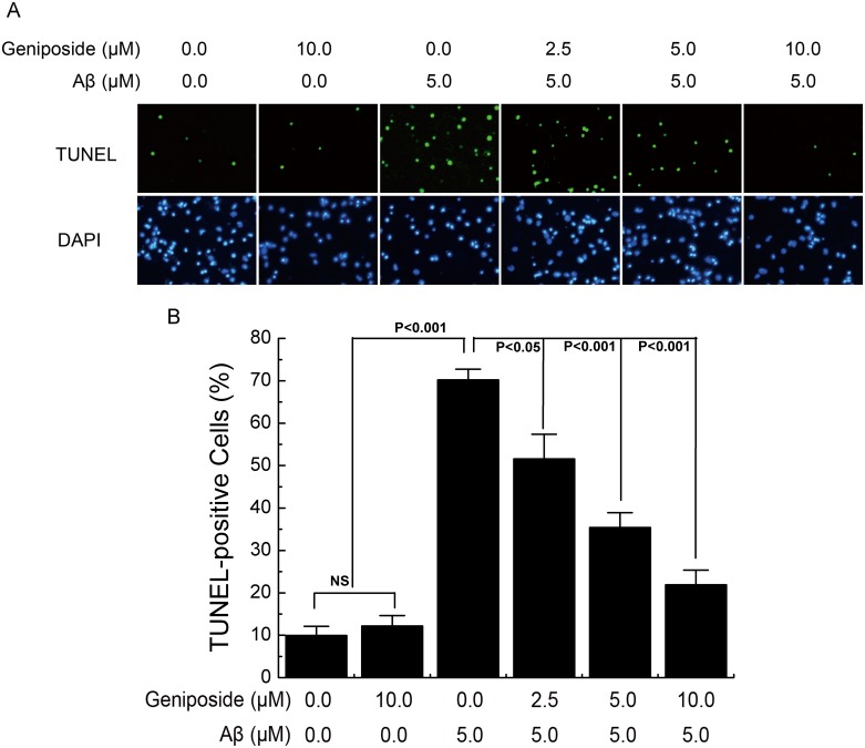
Fig 4. The effect of geniposide on Aβ1-42-induced apoptosis.
A, Photomicrographs of TUNEL and DAPI fluorescence staining. Neurons were cultured in the presence of 5 μM oligomeric Aβ1–42 and in the presence and absence of geniposide (2.5 μM, 5 μM, 10 μM) for 24 h. TUNEL-positive cells were stained in green while all cells’ nuclei were stained in blue. B, Quantitative analysis the ratio of TUNEL-positive cells to total neuron number. The ratio of TUNEL-labelled neurons was significantly increased in neurons treated with oligomeric Aβ1–42 for 24 hours while the geniposide treatment dramatically reduced the Aβ-induced TUNEL-positive cells in a dose-dependent manner. NS: non significance. N = 6 per group of cells. Studies were repeated thrice and data were expressed as mean ± SEM of percentage of vehicle-treated cells.

