Figure 2.
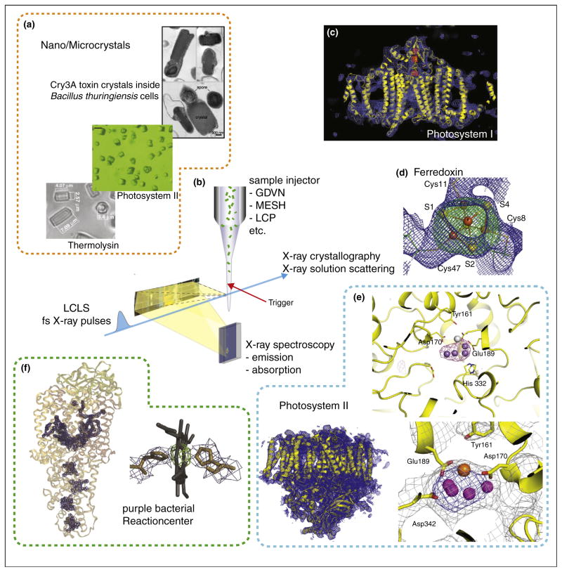
Jet based crystallography experiments of metalloenzymes at XFEL sources. (a) Examples of nano- to micrometer size crystals used, e.g. thermolysin [17], PS II [36] and toxin crystals (grown inside cells) [29]. (b) Schematic of the experimental approach using a liquid jet for sample delivery to the interaction point and collecting the forward scattering is shown. This setup can be combined with triggering by optical laser pulses and with spectroscopic measurements, e.g. X-ray emission spectroscopy. XFEL structures obtained: (c) PS I with electron density shown in blue and omit maps for the Fe4S4 clusters in red [25]. (d) Electron density for one of the Fe4S4 clusters in ferredoxin, with omit map shown in green [32]. (e) PS II with overall structure of one monomer (bottom left), the oxygen evolving complex (bottom right) [36] and the anomalous difference density from the Mn atoms in the OEC (top right) [31]. (f) PBRC with overall structure and omit maps for the cofactors (left) and detail for the electron density obtained for one of the heme groups (right) [33].

