Fig. 1.
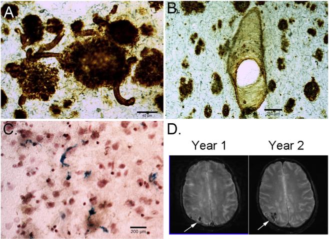
Cerebrovascular neuropathology in DS. (A) Beta-amyloid 1-42 immunostaining of the frontal cortex in 67-year old adult with DS and AD shows plaques and significant CAA in multiple small vessels (arrows). (B) Beta-amyloid 1-40 shows a different pattern with fewer plaques being labeled with CAA appearing more prominent, particularly in vessels (arrows). (C) Prussian blue staining of a 58-year old with DS and AD shows significant numbers of microhemorrhages (blue). T2* weighted MR images in a 60-year old male with DS, who is currently nondemented over a 2-year time interval, shows significant CAA in the occipital cortex that is progressively getting worse (white arrows) (images courtesy of Dr. David Powell, University of Kentucky).

