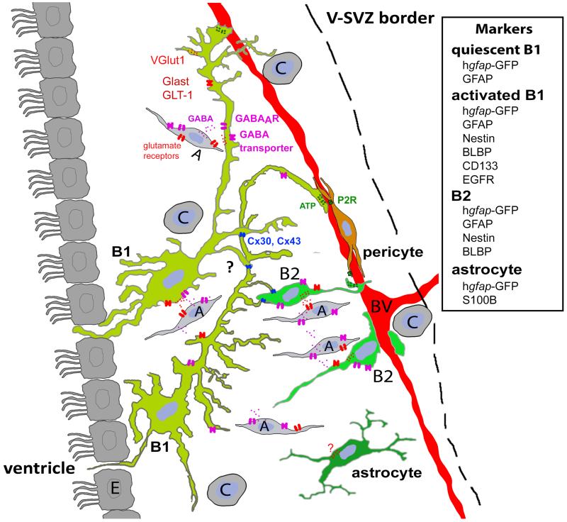
Figure 1.
The different populations of astrocyte like cells and their markers in the adult V-SVZ drawn from cells expressing GFP after electroporation.
Ependymal cells (E) are multiciliated located at the border of the lateral ventricle. Activated and quiescent type B1 cells extend an apical process intercalated between ependymal cells while their basal process contact the blood vessels. Type B2 cells, also called niche astrocytes, display an intermediate fusiform/stellate appearance and are located mostly at the periphery of the V-SVZ. They never contact the ventricle but interact with blood vessels. A third very discreet population of astrocyte was observed in the V-SVZ and is S100B positive. These cells present a stellate appearance (Platel et al. 2009) but their arborisation is not well developped. It is unknown if they connect to blood vessels. Type A cell (neuroblasts) have a migratory morphology and are located throughout the V-SVZ. Type C cells (transit amplifying progenitors) are located throughout the V-SVZ and apposed to blood vessels. Pericytes are located on capillaries and present several processes that run on the blood vessel.

