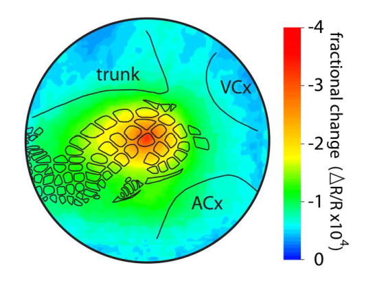
Fig. 1.

Neuronal activity spreads horizontally for long distances following single whisker stimulation. The intrinsic signal optical imaging response following stimulation of the C2 whisker (the first 500 msec containing the maximal areal extent of the initial dip activity) was averaged across 37 rats as described by Chen-Bee et al. (2012) and was plotted as a false-color image of fractional change relative to prestimulus values. The outer circle has a diameter of 7 mm and represents an extrapolation of the farthest electrode used in the recordings of Frostig et al. (2008), at which an evoked field potential response could be detected in 100% of animals. Black outlines show locations of whisker barrels and cytoarchitectonic areas that were detected by cytochrome oxidase staining in a representative animal. Trunk, trunk region of primary somatosensory cortex; VCx, visual cortex; ACx, auditory cortex
