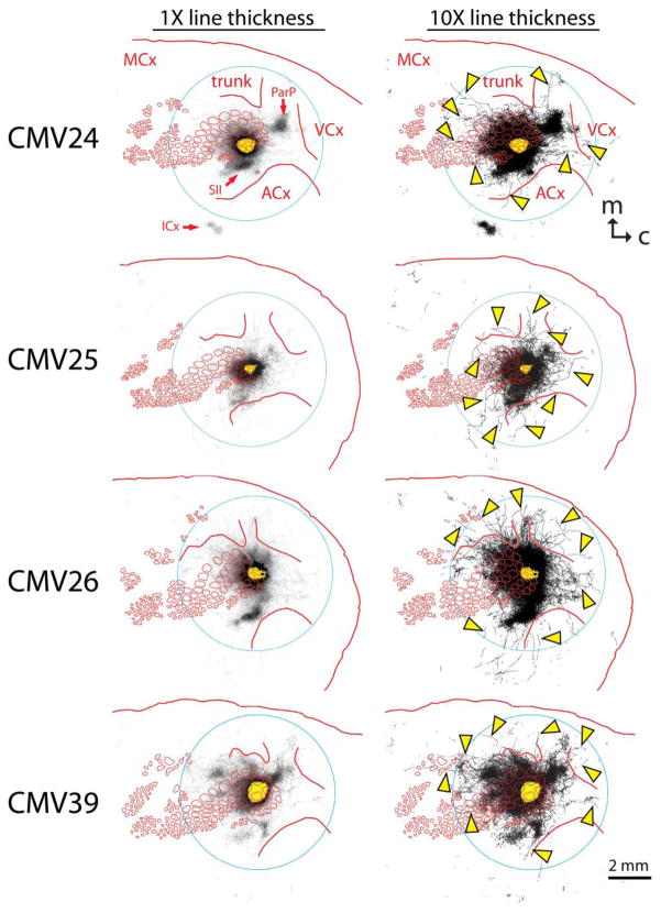
Fig. 2.
Labeled axons detected in the nine most shallow 40-μm sections from four brains injected with an AAV vector directing expression of GFP by way of a non-selective CMV promoter. Red lines indicate the locations of barrels and other cortical regions that were detected by staining layer 4 sections from the same brain for cytochrome oxidase activity. Left panels show tracings rendered using thin lines in order to appreciate the axons found within known specific projection areas including posterior parietal cortex (ParP), secondary somatosensory cortex (SII), insular cortex (ICx), and motor cortex (MCx). Right panels show tracings rendered using thicker lines in order to appreciate more sparsely distributed axons radiating in nearly all directions from the injection site. Several of the longer examples of these axons are indicated with arrowheads. Some of these axons crossed through dysgranular regions of cortex to penetrate the trunk region of somatosensory cortex (trunk), the visual cortex (VCx), and the auditory cortex (ACx). The yellow area in the center of each panel indicates the saturated immunostaining present in a section near layer 4, representing where the majority of the neurons infected by the virus were located (i.e., the “size” of the injection). This figure represents only a sampling of the axons that could have been reconstructed in these brains. Caudal (c) is to the right, and medial (m) is towards the top. The circles (7.2 mm in diameter) represent the area used for quantitative analysis in this study

