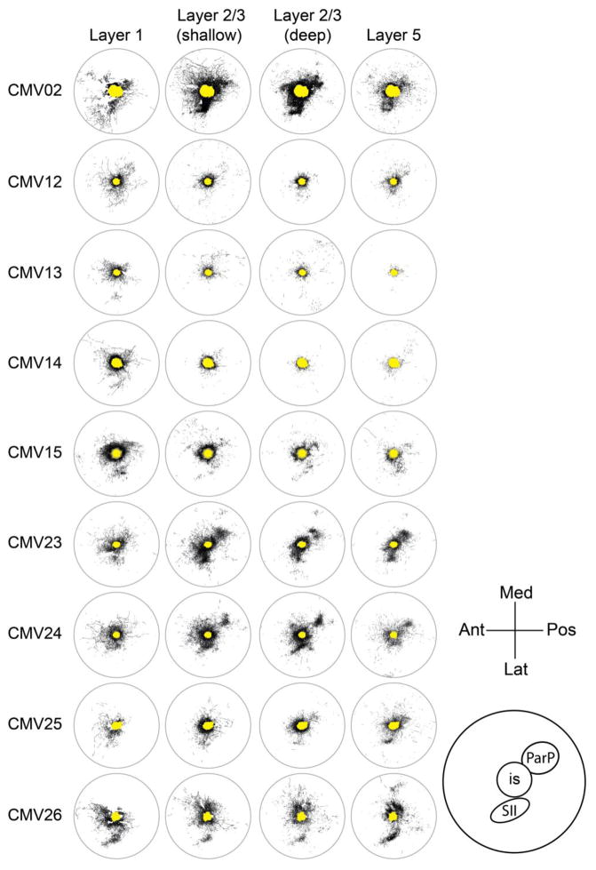
Fig. 3.
Tracings of axons within the 7.2-mm diameter analysis region of four sections from each of nine brains injected supragranularly with AAV-CMV-GFP. The similarity of axonal distributions across different depths in a given brain can be appreciated from this figure, as well as the differences in the absolute levels of labeling across different brains. The yellow area in the center of each tracing represents the extent of the saturated labeling at the core of the injection site in deep layer 2/3. Med, medial; Pos, posterior; Lat, lateral; Ant, anterior; SII, secondary somatosensory cortex; ParP, posterior parietal cortex; is, injection site

