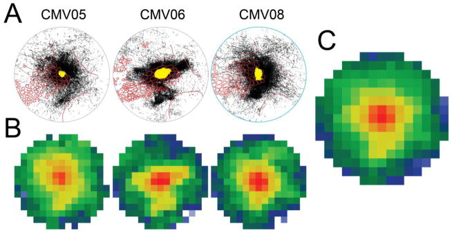
Fig. 8.
Projection patterns following infragranular injection of AAV-CMV-GFP. A. Tracings within the 7.2-mm diameter circular analysis region at four section depths were collapsed together for each of three brains injected infragranularly with AAV-CMV-GFP. These patterns can be compared with the patterns from supragranular injections shown in Fig. 4. The yellow area in the center of each tracing represents the extent of the saturated labeling at the core of the injection site in deep layer 2/3. B. Axonal densities resulting from the three infragranular injections were quantified as described for supragranular injections, averaged across section depth, and re-expressed as in the bottom row of Fig. 6. C. The re-expressed data arrays illustrated in B were averaged to reveal a largely symmetrical axonal radiation, similar to that found for supragranular injections (Fig. 7)

