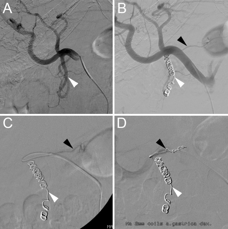
Fig. 3.
Typical angiography in a patient who underwent coil-embolization of the gastroduodenal artery (GDA) and right gastric artery (RGA). A Digital subtraction angiography (DSA) of the GDA (white arrowhead) on pre-treatment angiography. B DSA with appearance of the RGA (black arrowhead) after coil-embolization of the GDA. C DSA with catheter placement in the RGA. D DSA after successful coil-embolization of the GDA and RGA

