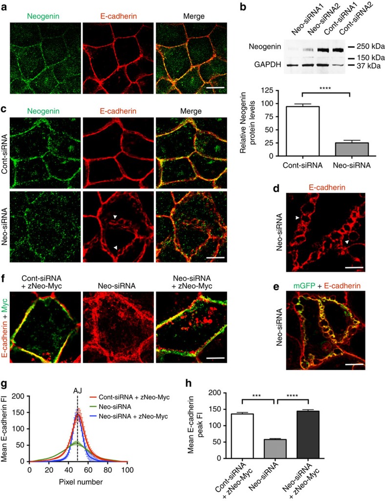
Figure 1. Neogenin maintains AJ integrity.
(a) Representative confocal micrograph showing apical view of Caco-2 cells where Neogenin (green) co-localized with Ecad (red; merge, yellow) at the AJ. (b) Immunoblot densitometry of Neogenin protein levels in Neo-siRNA cells relative to the control protein, GAPDH (n=3, mean±s.e.m., ****P<0.0001, Student's t-test). (c) Neo-siRNA induced the loss of adhesion between Ecad+ membranes, leading to the formation of tightly packed bleb-like structures at the AJ (arrowheads; Ecad, red; Neogenin, green). (d) Higher-magnification image of disrupted AJs in Neo-siRNA-expressing cells. (e) Co-localization of membrane-bound GFP (mGFP, green) and Ecad (red; merge, yellow) in Neo-siRNA-transfected cells. (f) Full-length zebrafish Neogenin (zNeo-Myc) rescued junctional integrity (Myc, green; Ecad, red). Line-scan analysis: (g) mean Ecad fluorescence intensity (FI) across AJs. (h) Mean Ecad peak FI at the AJ (n=150 junctions, mean±s.e.m., ***P<0.001, ****P<0.0001, one-way analysis of variance (ANOVA), Dunn's post hoc test). Scale bars, 15 μm (a,c); 5 μm (d,e); 7 μm (f).

