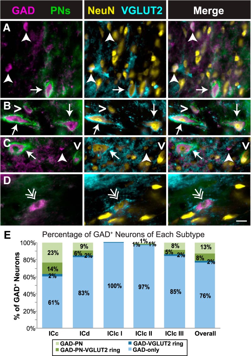
Figure 6.

GAD+ neurons can be divided into four distinct subtypes. A–D, Photographs of four subtypes of GAD+ neurons in the IC. Each image row shows a single field that was imaged to reveal four markers. The first column shows an overlay of images stained for GAD and PNs; the second column shows an overlay of images stained for NeuN and VGLUT2; the third column shows an overlay of all four images. Some neurons have both a PN and a ring of VGLUT2+ terminals (GAD–PN–VGLUT2 ring; arrows in A–C), some neurons only have a PN (GAD–PN; open arrowheads in B and C), some neurons only have a ring of VGLUT2+ terminals (GAD–VGLUT2 ring; double arrow in D), and some neurons lack both a PN and a ring of VGLUT2+ terminals (GAD-only; arrowheads in A and C). The complete lack of PN staining in D is attributable to a local lack of PNs rather than a staining issue, because other photographs (not shown here) of PN-surrounded cells were collected from the same section, and a small amount of WFA labeling of the extracellular matrix is present in the background. Scale bar, 20 μm. E, Bar graph showing the proportion of each GAD subtype in each IC subdivision. GAD+ neurons lacking both a PN and a ring of VGLUT2+ terminals (light blue) are the majority in each subdivision but are a smaller majority in ICc. Overall and in each subdivision, most GAD+ neurons with a PN (light and dark green bars) lack a VGLUT2 ring (light green bars).
