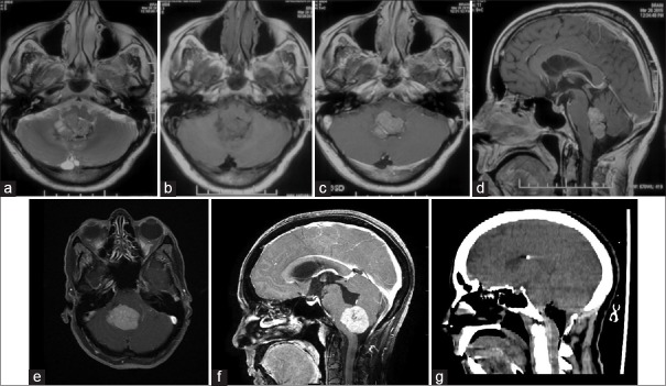
Figure 1.
(a) T2-weighted image showing a heterogenous isointense intra fourth ventricular mass, (b) the lesion is isointense on T1-weighted imaging, (c and d) the lesion is enhancing on contrast administration, and it has multiple lobulations and is entirely inside the fourth ventricle. (e and f) contrast enhanced axial magnetic resonance imaging of and sagittal computed tomography, respectively of case 2 and (g) a postoperative computed tomography showing complete excision

