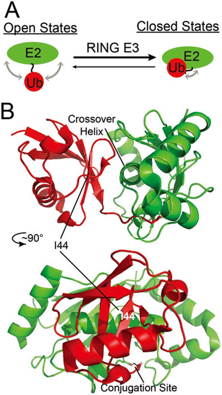
Figure 2.
Ubiquitin positioning in the E2∼Ub conjugate. (A) A cartoon depiction of the dynamic “open states” in which Ub samples a range of various positions relative to the E2, but shifts towards population of more “closed states” upon binding of a RING-type E3. (B) Crystal structure of an E2∼Ub oxyester conjugate (Ube2D2) in a closed state showing the interface formed between the E2 (green) crossover helix and the I44 surface of Ub (red) upon binding a RING E3 (BIRC7 (not shown); PDBID: 4AUQ). The two representations are the same structure rotated ∼90° about the vertical axis. The E2 is in the same orientation as in Figure 1 in the bottom representation.

