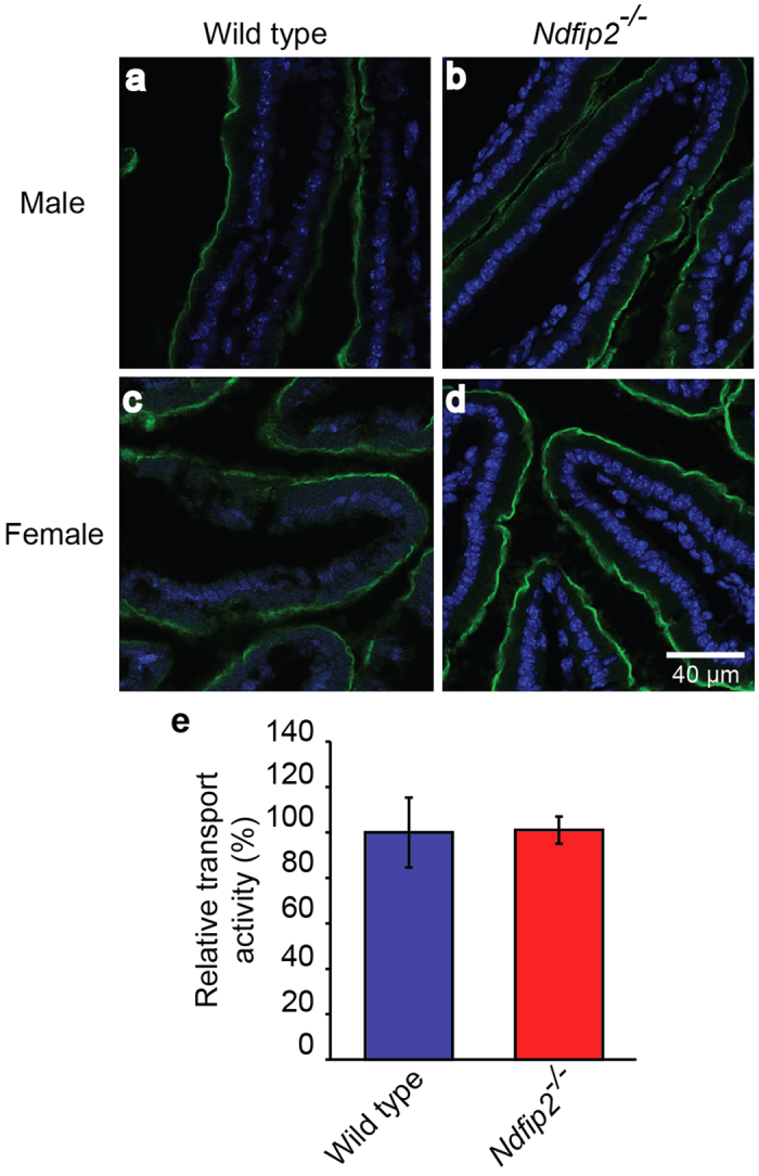
Figure 2. Ndfip2-deficient mice show normal DMT1 expression and activity.

DMT1 expression on the apical surface of the duodenum (green) of (a) male wild type and (b) Ndfip2−/− mice, and (c) female wild type and (d) Ndfip2−/− mice fed a low iron diet for three weeks. Representative images from n = 4. Blue shows DAPI staining of the nuclei. (e) DMT1 relative transport activity as measured by the fluorescence quenching assay in enterocytes isolated from wild type and Ndfip2−/− mice fed a low iron diet show no significant difference. Data represented as mean ± s.e.m.; n = 5.
