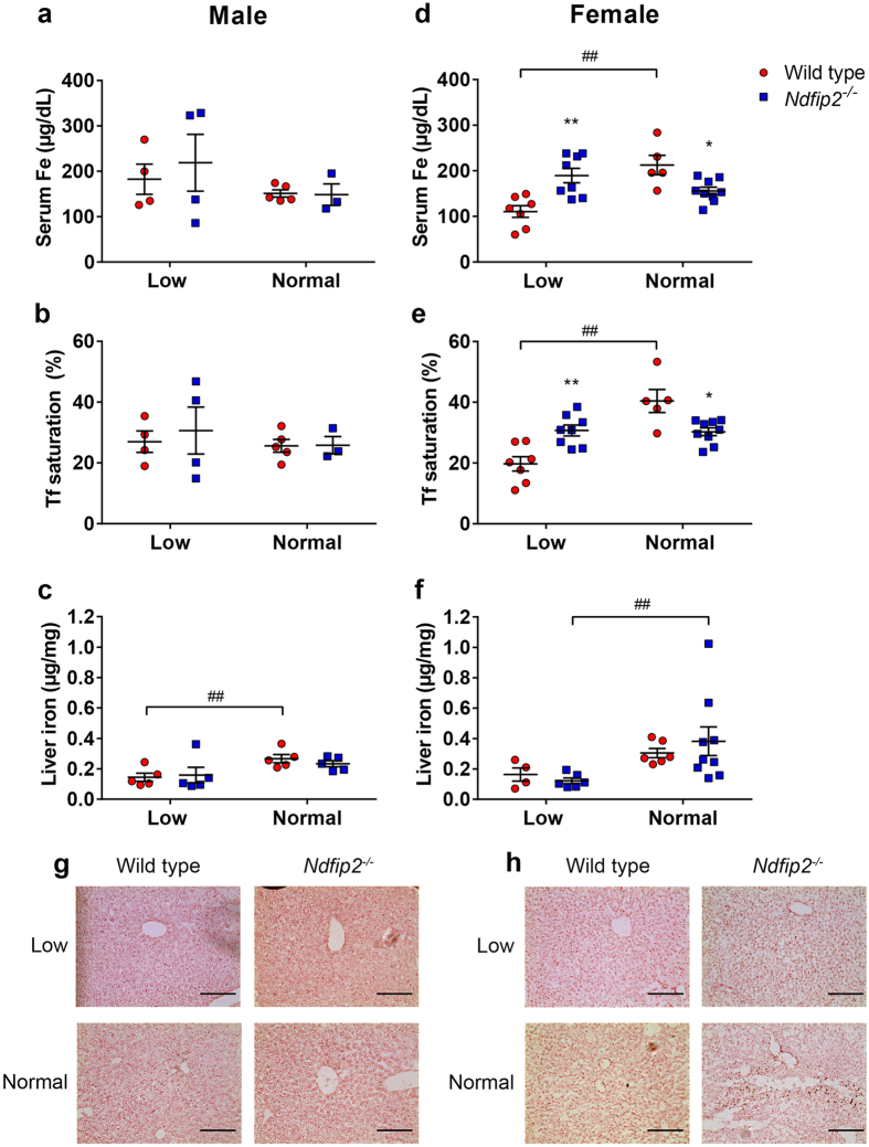
Figure 3. Sex-specific changes in serum iron, transferrin saturation and liver iron measurements in mice fed a low or normal iron diet.
(a,d) Serum iron levels and (b,e) transferrin (Tf) saturation in male and female wild type and Ndfip2−/− mice fed a normal or low iron diet. Male mice show no significant differences in serum iron or Tf saturation between normal and low iron diet in either wild type or knockout mice. Wild type female mice show a significant decrease in serum iron (##p < 0.0001) and Tf saturation (##p < 0.0001) when fed a low iron diet, but Ndfip2−/−females are able to maintain their levels when challenged. *represents a significant difference in serum iron (p = 0.0232) and Tf saturation (p = 0.008) between wild type and knockout females fed a normal diet. **represents a significant difference in serum iron (p = 0.0008) and Tf saturation (p = 0.0021) between wild type and knockout females fed a low iron diet. (c,f) Liver iron levels in wild type and Ndfip2−/−mice fed a normal or low iron diet. Wild type males show a significant reduction in liver iron levels when fed a low iron diet (##p = 0.0425), but Ndfip2−/−males are able to maintain liver iron levels. Ndfip2−/− females show a decrease in liver iron levels (##p = 0.0276). Red circles, wild type; blue squares, Ndfip2−/−. Data represent mean ± s.e.m.; n = 4–8. (g) Liver iron levels in male and (h) female mice as measured by Perl’s Prussian blue staining. Ndfip2−/− female mice fed a normal diet show significant iron deposits compared to wild type mice, and male mice do not show any significant staining.

