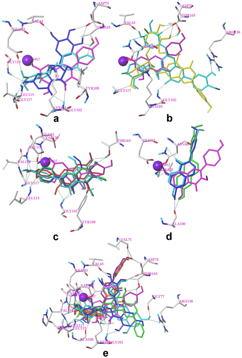
Figure 5. The predicted binding modes of 17 flavonoids to GyrB.
Black dashed lines represent H-bonds between the flavonoids and the protein active site residues. Magnesium atom was shown in purple ball. The binding mode of compound 14 (magenta) was displayed for comparison. (a) 18(blue) and 19 (cyan); (b) 20(yellow) and 22(cyan), two glycosidic groups superposed well to each other; (c) 1(cyan), 2(green), 3(red) and 4(blue); (d) 23(cyan), 26(green) and 27(blue); (e) 10(green), 16(blue), 24(red), 25(purple), 28(orange) and 30(cyan), the glycosidic or gallate group superposed well to each other for all the flavonoids.

