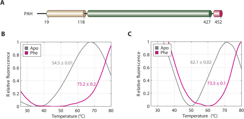
Figure 1. Interaction between PAH-RD and Phenylalanine.
(A) Schematic of the regulatory domain (brown), catalytic domain (green) and tetramerization domain (pink) of the human PAH polypeptide. (B) DSF of the unliganded (grey line; Tm = 54.5 °C ± 0.01 SD) and Phe-bound (pink line; Tm = 75.2 °C ± 0.2 SD) hPAH-RD1–118. (C) DSF of the unliganded (grey line; Tm = 62.1 °C ± 0.02 SD) and Phe-bound (pink line; Tm = 73.3 °C ± 0.1 SD) hPAH-RD19–118.

