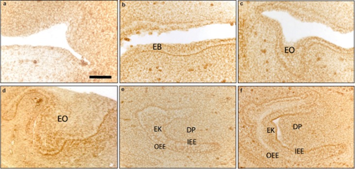
Figure 2.
The expression pattern of Hsp 60 in early stages of lower incisor development in mice. (a,b) Hsp 60 is present in the oral epithelium and EB in the initial stage of the tooth development . (c–f) From E12.5 to E15.5, Hsp 60 protein is present in the structures of the developing enamel organ (EO). From E14.5, the IEE, OEE and EK of the enamel organ were positive for Hsp 60. The cells of DP show weak Hsp 60 expression. a, E10.5; b, E11.5; c, E12.5; d, E13.5; e, E14.5; f, E15.5. Scale bar: a–d, 50 µm; e and f, 100 µm. DP, dental papilla; EB, epithelialband; band; EK, enamel knot; EO, enamel organ; IEE, inner enamel epithelium; OEE, outer enamel epithelium.

