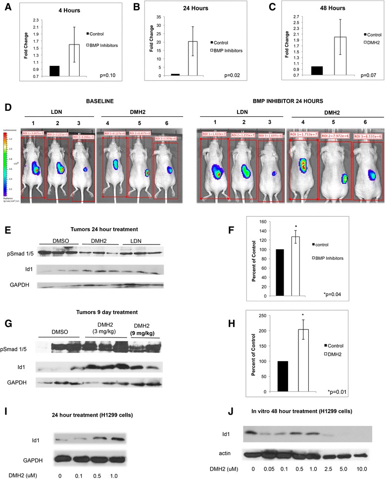
Fig. 1.

DMH2 causes an increase in Id1 expression in tumor xenografts and in vitro at low concentrations. a Mice with established tumors from H1299 Id-luc cells were treated with DMSO or 3 mg/kg of DMH2 or 3 mg/kg LDN and after 4 h tumor luminescence determined and compared to baseline (n = 3 for each group). (b) Two days later mice were treated with DMSO or 3 mg/kg of DMH2 or 3 mg/kg LDN every 8 h for 24 h and tumor luminescence determined and compared to baseline. (c) In a separate experiment, established tumors were treated with DMSO (n = 3) or 3 mg/kg of DMH2 (n = 5) twice daily for 48 h and luminescence was compared to baseline. (d) Bioluminescence images of H1299 Id1-luc tumors before and after treatment with BMP inhibitors for 24 h. Data depicted as fold increase in tumor luminescence of mice treated with inhibitors compared to baseline. (e) Western blot analysis of tumor xenografts from mice treated with DMSO or 3 mg/kg of DMH2 or 3 mg/kg LDN every 8 h for 24 h and (f) the mean optical density readings of Id1 from the corresponding Western blot was normalized to GAPDH and presented as percent of control. (g) Western blot analysis of xenografts from mice treated with twice-daily injection of DMH2 for 9 days and (h) the mean optical density readings of Id1 from the corresponding Western blot was normalized to GAPDH and presented as percent of control. (i, j) Western blot analysis of H1299 cells in cell culture treated with increasing doses of DMH2 for 24 (n = 2) (i) and (j) 48 h (n = 4)
