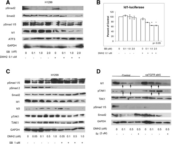
Fig. 4.

Inhibition of both BMP and TGFβ signaling enhances the downregulation of pTAK1 and Id1. a Western blot analysis of H1299 cells treated with increasing doses of the TGFβ inhibitor SB-505124 (SB) with and without DMH2. SB and DMH2 together enhanced the downregulation of Id1 (n = 3). (b) Id1-luciferase assay demonstrating decreased Id1 promoter activity only in cells treated with both SB and DMH2 (n = 2). (c) Western blot analysis of H1299 cells treated with 1 μM SB and increasing doses of DMH2 (n = 4). The combination of SB and DMH2 enhanced the downregulation of Id1, Id3, and pTAK1. (d) H1299 cells were transfected with constitutively active alk5 (ca alk5) or empty vector and treated with DMSO or DMH2 for 48 h (n = 3). Cells were also treated with or without 2 μM 5z for 24 h. Western blot shows that when BMP signaling is inhibited caAlk5 increases Id1 and pTAK1 expression that is attenuated with 5z. Arrows show increased expression of Id1 and pTAK1 in cells treated with caAlk5 and DMH2 compared to controls
