
Fig. 3.
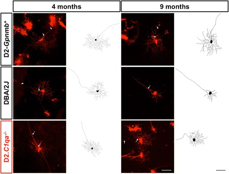
Representative DiOlistic and tracings of mouse retinal ganglion cells. Retinal ganglion cells from D2.Gpnmb +, DBA/2 J and D2.C1qa -/- retinas were DiOlistically labelled to analyse dendritic morphology. Top row: representative images and tracings from 4 month (left) and 9 month (right) D2.Gpnmb + retinal ganglion cells. Centre row: representative images and tracings from 4 month (left) and 9 month (right) DBA/2 J retinal ganglion cells. Bottom row: representative images and tracings from 4 month (left) and 9 month (right) D2.C1qa -/- retinal ganglion cells. Scale bar = 100 μm, white arrows = axon