Fig. 6.

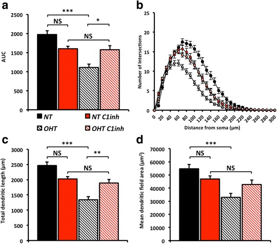
Inhibition of C1 prevents retinal ganglion cell dendrite pruning in ocular hypertensive rat eyes. To test dendrite loss in the rat bead model of glaucoma we DiOlistically labelled retinal ganglion cells following an ocular hypertensive insult. There was a dramatic decrease in dendritic integrity of retinal ganglion cells in ocular hypertensive eyes compared with normotensive eyes as assessed by Sholl analysis (a, area under the curve (AUC), b, Sholl analysis). This is paralleled in both the retinal ganglion cell dendritic field area (c) and total dendritic length (d). NT = normotensive eyes, sham control, OHT = ocular hypertensive eyes. Error bars = SEM, * = P < 0.05, ** = P < 0.01, *** = P < 0.001
