
Fig. 7.
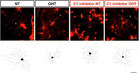
Representative DiOlistic and tracings of rat retinal ganglion cells. Retinal ganglion cells from normotensive eyes and ocular hypertensive eyes were DiOlistically labelled to analyse dendritic morphology. Top row: representative images and Bottom row: tracings from normotensive (far left), ocular hypertensive (centre left) normotensive (centre right), and ocular hypertensive (far right) retinal ganglion cells treated with C1 inhibitor. Scale bar = 100 μm, white arrows = axon