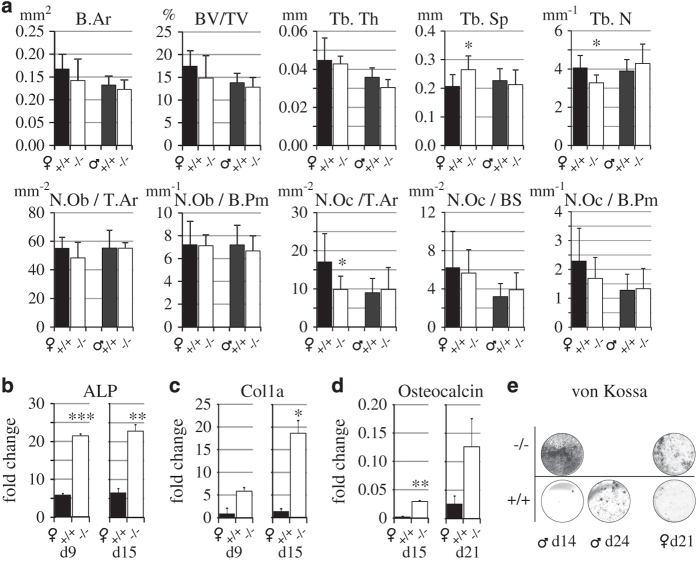
Figure 5.
Histomorphometry of tibial metaphysis and osteogenic differentiation of bone marrow cells. (a) Histomorphometric analysis of tibial metaphysis of 3-month-old WT (+/+, black bars) and KO (−/−, open bars) female and male mice. The only significant difference was the reduced osteoclast number in female mice. N=8 per group. RT-qPCR analysis of (b) ALP, (c) collagen 1a and (c) osteocalcin during osteogenic differentiation of bone marrow-derived MSCs of 8-week-old female WT and KO mice. (e) Von Kossa staining of a separate set of male and female KO and WT osteogenic cell cultures. KO cultures showed strong mineralization 1 week earlier that WT cultures. The brightness and contrast of the images were brought to visual uniformity. Similar results as shown in b, c, d and e were obtained from three independent experiments.

