
Figure 1. Hippocampal-specific ATF4 down-regulation.
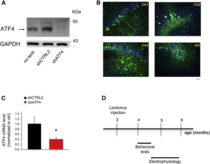
(A) Representative western blot showing ATF4 protein knockdown in primary hippocampal neuronal cultures infected at 5 DIV for 2 weeks with shATF4-lentivirus compared to shCTRL2-infected and non-infected cultures.
(B) Distribution and expression of GFP in mouse hippocampus 1 month after infection with GFP-expressing lentivirus. Sections of lentivirus-injected animals were immunolabeled with antibody against GFP (green) and counterstained with Topro3 (nuclei, blue). Scale bar 20μm.
(C) RT-qPCR analysis of ATF4 mRNA levels in mouse hippocampus 4 weeks after lenti-shATF4 injection. Data are expressed as mean ± SEM (shCTRL2 n=7, shATF4 n=7) (*p<0.05).
(D) Scheme of experimental design: 3-month-old hippocampi of C57BL/6 wt male mice were stereotaxically inoculated either with lenti-shCTRL2, -shCTRL1, or -shATF4. 4 weeks after injection, cognitive functions of the animals were assessed with a battery of behavioral tests, at the end of which they were sacrificed for electrophysiological analyses.