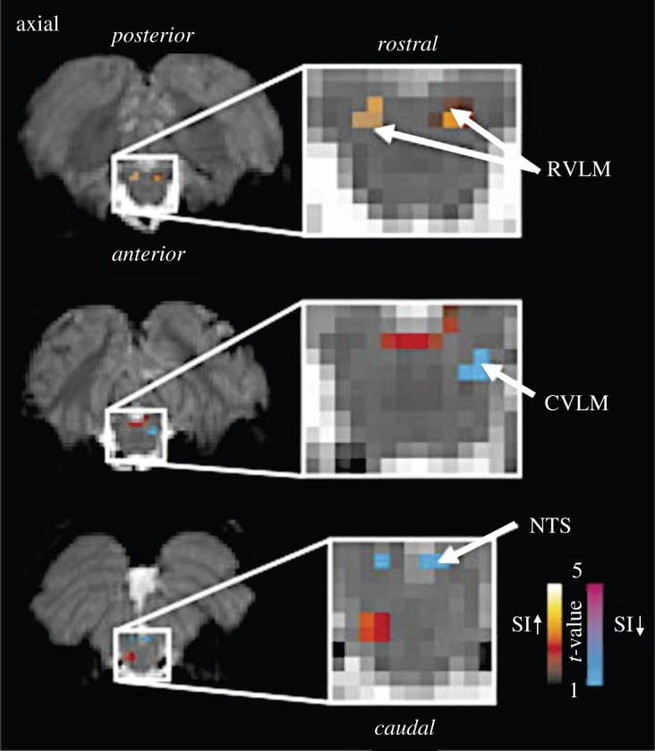
Figure 2.

fMRI signal intensity changes correlated with sympathetic outflow. fMRI signal intensity changes correlated with spontaneous fluctuations in muscle sympathetic nerve activity (MSNA) in eight subjects. Increases (hot colour scale) and decreases(cool colour scale) in fMRI signal intensity with increases in MSNA are overlaid onto a series of axial fMRI slices from an individual subject. RVLM, rostral ventrolateral medulla; NTS, nucleus tractus solitarius; CVLM, caudal ventrolateral medulla; SI, signal intensity. Adapted from [58]. (Online version in colour.)
