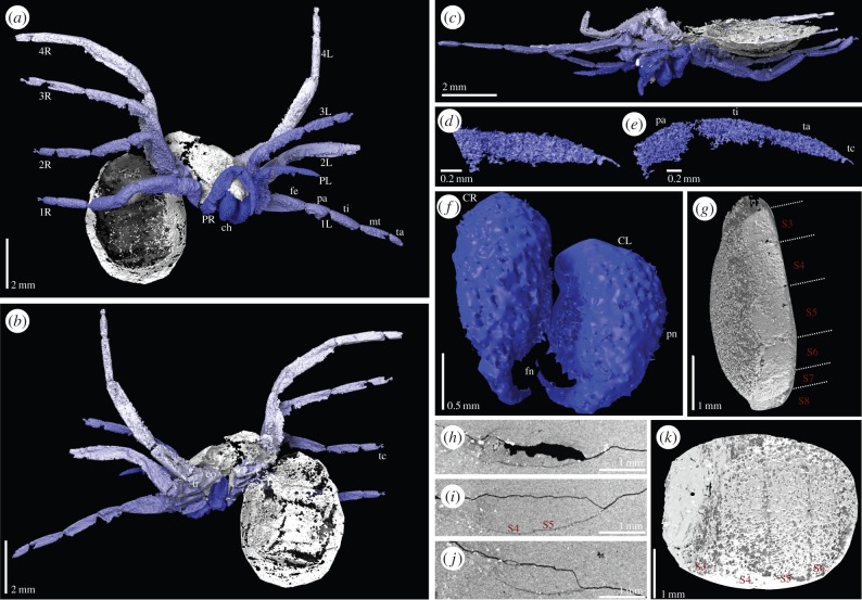
Figure 2.
Digital visualization of Idmonarachne brasieri gen. et sp. nov. based on laboratory and synchrotron scans of the fossil. (a–c) Laboratory-based scans. (a) Prosoma in anterior view, and ventral opisthosoma of the specimen, with chelicerae tucked between pedipalps, ventral to the clypeus. (b) Dorsal opisthosoma, and prosoma in posterior view, showing some opisthosomal segmentation. (c) Ventral view of prosoma, leg coxae and cheliceral termination apparent. (d–k) Synchrotron scans. (d–e) Tips of pedipalps showing claw and onychium. (f) Isolated chelicerae in detail, comprising paturon and fang. (g) Lateral view of ventral opisthosoma, ventral plates numbered as described in the text—fourth and fifth lacking spinnerets. (h)–(j) Computed slice images showing the opisthosoma in cross section, posterior right, ventral bottom. Sternal plates are preserved as thin, but continuous pieces of cuticle. (k) Ventral view of opisthosoma, ventral plates numbered—fourth and fifth lacking spinnerets. 1L–4L, first to fourth left leg; 1R–4R, first to fourth right leg; ch, chelicerae; CL, left chelicera; CR, right chelicera; co, coxa; fe, femur; fn, fang; pa, patella; PL, left pedipalp; pn, paturon; PR, right pedipalp; S3–S9, ventral plates 3–9; tc, terminal claw; ti, tibia; tr, trochanter.

