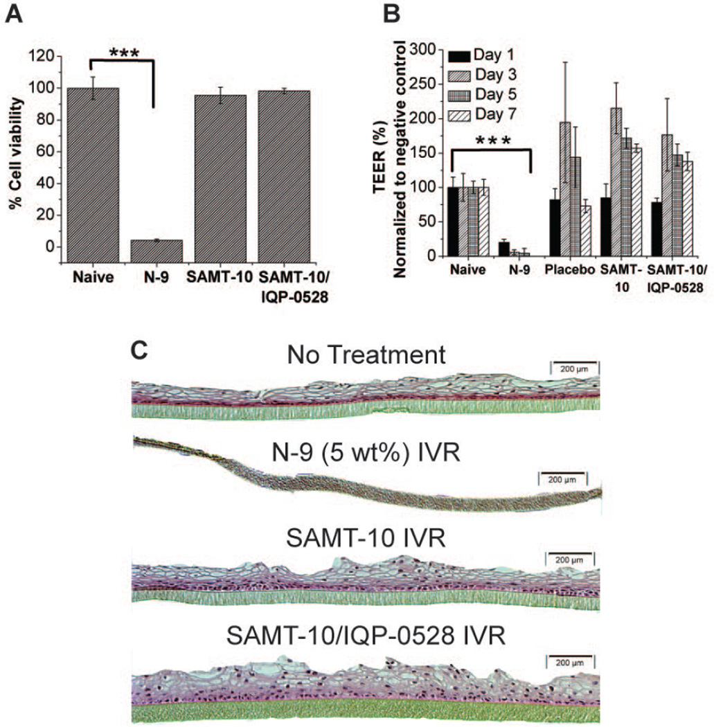
Figure 6.
(A) VEC-100 tissue viability in the MTT assay after seven days of treatment with the SAMT-10 IVR (8 wt%) and the SAMT-10/IQP-0528 combination IVR. The percent viability response was compared with the naïve samples (no treatment tissues). There was no statistically significant difference between the tested IVRs and the naïve tissues, but the N-9-treated tissues showed significantly lower viability (*statistical significance, p < 0.05, two-tailed, Student’s t test). (B) Percent TEER on days 1, 3, 5 and 7 for the control (naïve), the N-9 treated IVR segment, the placebo IVR segment and the drug-loaded IVRs. The percent TEER for the N-9 treated tissues was significantly lower than that of the naïve tissues on all of the test days, but no significant loss in TEER was observed for the ARV-loaded formulations. (C) H&E stained images of naïve tissue, N-9-dosed tissue, SAMT-10 IVR treated tissue and SAMT-10/IQP-0528 IVR treated tissue after seven days of exposure (10X magnification). There was a complete loss of the epithelium in the N-9 dosed tissues, which indicated toxicity, whereas no epithelial erosion was observed in the drug-loaded IVR tissues.

