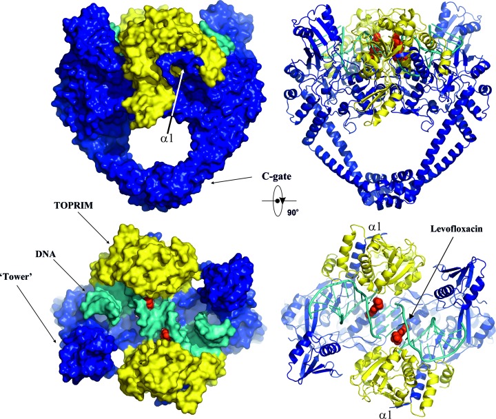
Figure 3.
Overall orthogonal views of the cleavage complex of topoisomerase IV from K. pneumoniae in surface (left) and cartoon (right) representations. The ParC subunit is in blue, ParE is in yellow and DNA is in cyan. The bound quinolone molecules (levofloxacin) are in red and are shown using van der Waals representation.

