Fig. 1.
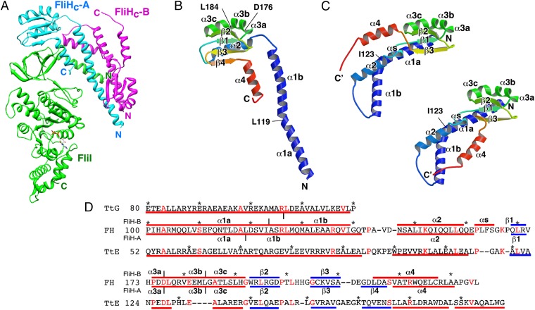
Structure of the FliHC2–FliI complex. (A) Cα ribbon drawing of the FliHC2–FliI complex. FliI and two FliHC subunits (FliHC-A and FliHC-B) are shown in green, cyan, and magenta, respectively. (B and C) Structure of FliHC. Cα ribbon representation of FliHC-A (B) and FliHC-B (C) shown in rainbow colors from the N terminus (blue) to the C terminus (red). The secondary structure elements are labeled. (C) FliHC-B shows two distinct conformations in the dimeric unit. FliHC-B of trimer-1 (trimer-3) (Upper Left) and that of trimer-2 (trimer-4) (Lower Right) are shown. (D) Structure-based amino acid sequence alignment of FliHC and the E (TtE) and G (TtG) subunits of A-ATPase from T. thermophilus (PDB ID code 3V6I). The red and blue bars indicate α-helices and β-strands, respectively. The secondary structures of FliHC-A and FliHC-B are shown below and above the FliH sequence, respectively, with the labels of the secondary structure elements. Conserved residues are highlighted in red characters. Every 10 residues are denoted with asterisks.

