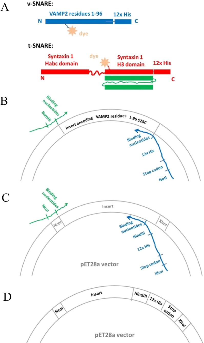
Fig. S5.
Protein constructs for the FRET/SFA studies. (A) Diagram of the design of the SNARE proteins, which contain two specific functional sites. The Cys residue within the SNARE domain is designed for site-specific fluorescence labeling, and the 12× His on the end of protein sequences provides the capability to anchor to a bilayer. (B) Illustration of the strategy of introducing the C-terminal 12× His to the v-SNARE through PCR cloning. (C) Illustration of the strategy of introducing the C-terminal 12× His to the pET28a vector through PCR cloning. (D) Illustration of the modified pET28a vector that contains C-terminal 12× His.

