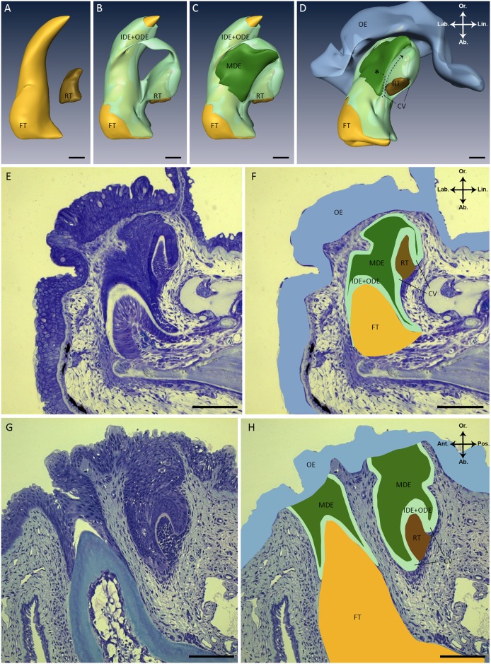
Fig 1. Structure of the dental organ in S. salar.
(A-D) Three-dimensional reconstructions of a tooth family on the lower jaw in a juvenile S. salar (fork length: 4.84 cm). To simplify, the inner dental epithelium (IDE) of the functional tooth and of its successor were combined with the outer dental epithelium (ODE) into one layer (Fig 1B to 1D, light green). Please note that the smoothened surface facing the observer represents the edge of the 3D reconstruction and does not indicate that the MDE lies superficially. Instead, the MDE is covered by IDE+ODE outside the area of reconstruction. (D) The same three-dimensional reconstruction as in (A-C) but slightly tilted. The hypothesis to be tested is that stem cells (asterisk) reside in the middle dental epithelium (MDE), and give rise to transit amplifying cells, populating the cervical loop to eventually become differentiated ameloblasts (dashed arrow). (E) Sagittal histological section (2μm) through a tooth family of a juvenile S. salar (fork length: 4.84 cm) stained with toluidine blue. These serial sections are used to generate the three-dimensional reconstructions in (A-D). (F) Same histological section as in (E) with different structures overlaid in the colors used in the three-dimensional reconstructions (A-D). (G) Transverse histological section (2μm) through two adjacent tooth families of juvenile S. salar (fork length: 10 cm). In one tooth family the replacement tooth is not visible and lies behind the plane of view, while in the other tooth family the functional tooth is not visible because it lies above the plane of view. (H) Same histological section as in (G) with different structures overlaid in the colors used in the three-dimensional reconstructions (A-D). Abbreviations: Ab: aboral; Ant: anterior; CV: cervical loop; FT: functional tooth; IDE: inner dental epithelium; Lab: labial; Lin: lingual; MDE: middle dental epithelium; OE: oral epithelium; ODE: outer dental epithelium; Or: oral; Pos.: posterior; RT: replacement tooth; asterisk: hypothetical stem cell niche; color codes: yellow (FT), brown (RT), light green (IDE+ODE), dark green (MDE), blue (OE); scale bars: 80 μm.

