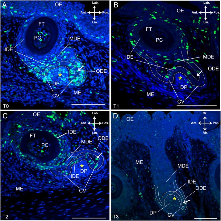
Fig 2. BrdU labelled tooth families in S. salar chased over different periods.
Immunohistological staining of frontal (A-C) and sagittal (D) sections through a tooth family on the lower jaw of S. salar with BrdU labelled cells in green and DAPI counterstain in blue. The tooth families presented are in a similar state of development: a young functional tooth and its successor in morphogenesis stage. (A) Pulsed specimen, T0; (B) Chase time T1, one week after BrdU administration; (C) Chase time T2, two weeks after BrdU administration and (D) Chase time T3, four weeks after BrdU administration. The white arrow indicates single cells within the middle dental epithelium (MDE) enclosed by the IDE, cervical loop and ODE of the replacement tooth. Abbreviations: Ab: aboral; Ant: anterior; CV: cervical loop; DP: dental papilla; FT: functional tooth; IDE: inner dental epithelium; Lab: labial; Lin: lingual; MDE: middle dental epithelium; ME: mesenchyme; OE: oral epithelium; ODE: outer dental epithelium; Or: oral; PC: pulp cavity; Pos: posterior; yellow asterisk: replacement tooth; scale bars: 100 μm.

