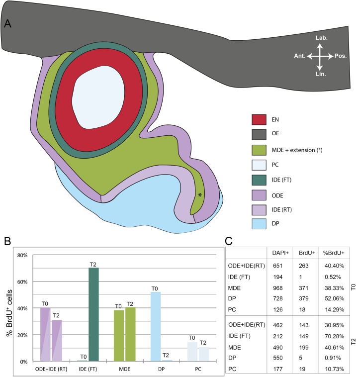
Fig 3. Ratio of BrdU+ to DAPI+ cells after pulse and after two weeks of chase time in S. salar.
(A) Schematic representation of the different layers in a tooth family containing two members: a young functional tooth and a successor in morphogenesis stage. (B) Graphical representation of the ratio of BrdU+ to DAPI+ cells in the different layers as depicted in (A). (C) Table showing results from cell counting in the different layers. The data were acquired from five consecutive sections in both T0 (pulsed specimen) and T2 (two weeks after BrdU administration). Abbreviations: Ant: anterior; DP: dental papilla; EN: enameloid; FT: functional tooth; IDE: inner dental epithelium; Lab: labial; Lin: lingual; MDE: middle dental epithelium; OE: oral epithelium; ODE: outer dental epithelium; PC: pulp cavity; Pos: posterior; RT: replacement tooth.

