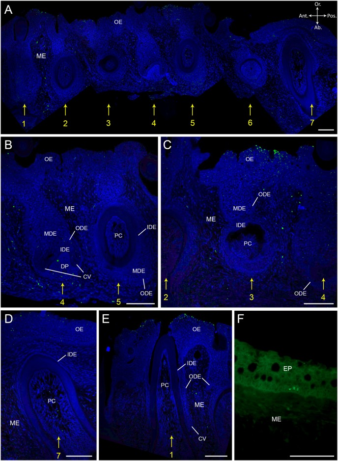
Fig 4. BrdU labelling after eight weeks of chase time in S. salar.
Immunohistological staining of sagittal sections through the lower jaw of S. salar having experienced a chase time of eight weeks (T4) with BrdU labelled cells in green and DAPI counterstain in blue. (A) shows seven adjacent tooth families in different stages of development. Tooth family one, four and seven have a young functional tooth and a replacement tooth in morphogenesis stage; tooth family two and five have an old tooth in resorption, a young successor, and show initiation of a third tooth germ; tooth family three and six have a mature functional tooth with its successor in cytodifferentiation stage. (B) In tooth family four, nuclei in the cervical loop, ODE and MDE of the successor show small fragments of BrdU label. Bright BrdU label is visible in the mesenchymal cells at the aboral side of the dental papilla. Label is absent in tooth family five. (C) Tooth family three, with a successor in cytodifferentiation stage, shows no BrdU labelled cells. Differentiated cells at the surface of the oral epithelium are brightly BrdU+. (D-E) show a young functional tooth of tooth family seven and one, resp., with fragmented BrdU label in the center region of the pulp cavity. (F) Skin epithelium with BrdU+ cells (LRCs) in the basal layer of the epithelium. Abbreviations: Ab: aboral; Ant: anterior; CV: cervical loop; DP: dental papilla; EP: skin epithelium; IDE: inner dental epithelium; MDE: middle dental epithelium; ME: mesenchyme; OE: oral epithelium; ODE: outer dental epithelium; Or: oral; PC: pulp cavity; Pos: posterior; asterisk: replacement tooth; yellow numbers indicate tooth family number; scale bars: 50 μm.

