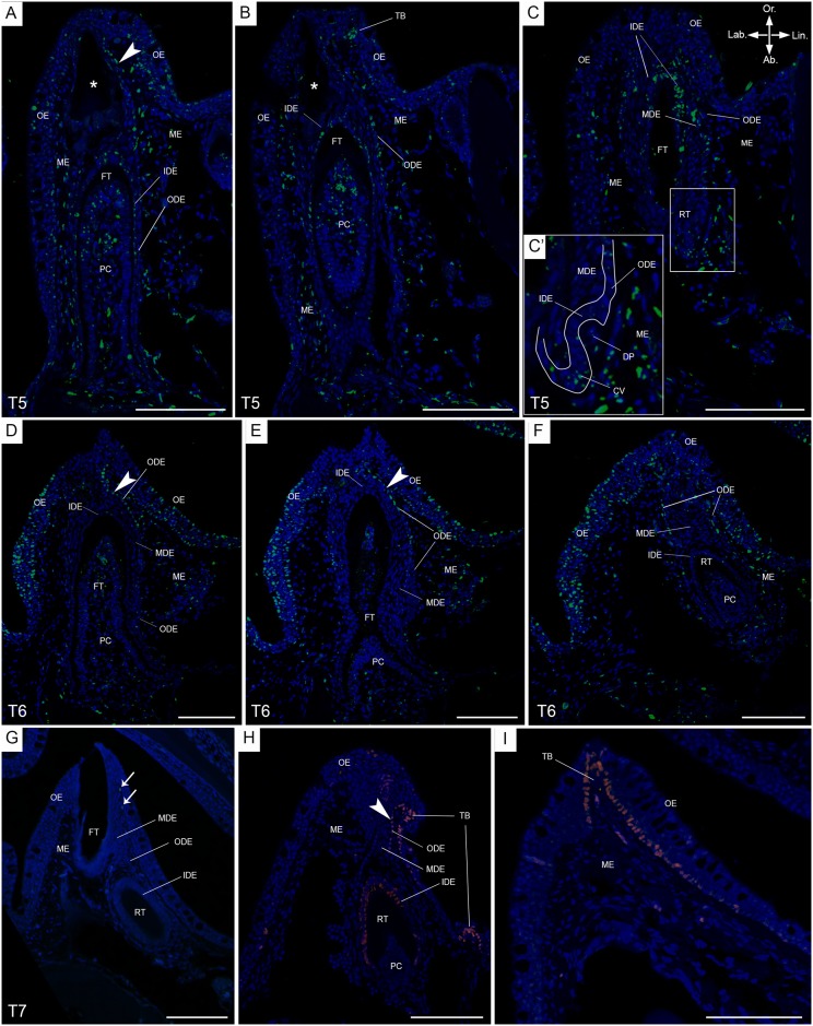
Fig 5. BrdU labelling after long chase times, and Sox2 distribution, in P. senegalus.
Immunohistological staining of transverse sections of a single tooth family on the lower jaw in P. senegalus after a chase time of six weeks (T5, A-C), eight weeks (T6, D-F) and twelve weeks (T7, G), with BrdU labelled cells in green and DAPI counterstain in blue. In A-C, (A) is the most anterior and (C) the most posterior section. (C’) is a magnification of the secondary successor, in morphogenesis stage. The anterior-posterior axis is perpendicular to the plane of sectioning, with the anterior side pointing upwards. In D-F, (D) is the most anterior and (F) the most posterior section. The anterior-posterior axis is perpendicular to the plane of sectioning, with the anterior side pointing upwards. This tooth family has a mature functional tooth and its successor in an advanced cytodifferentiation stage. (G) BrdU+ cells (LRCs) are present in the mesenchyme (arrows). (H-I) Distribution of Sox2 (pink) on transverse sections of a single tooth family (H) and taste bud (I); Sox2+ cells are distributed in the ODE transition zone, basal layer of the oral epithelium, IDE of a replacement tooth in cytodifferentiation stage and basal layer of the taste bud. Abbreviations: Ab: aboral; CV: cervical loop; DP: dental papilla; FT: functional tooth; IDE: inner dental epithelium; Lab: labial; Lin: lingual; MDE: middle dental epithelium; ME: mesenchyme; ODE: outer dental epithelium; OE: oral epithelium; Or: oral; PC: pulp cavity; RT: replacement tooth; TB: taste bud; arrowheads: ODE transition zone; asterisk: tooth in resorption; scale bars: 100 μm.

