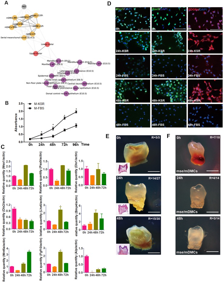
Fig 5. KSR, Fgf2 and Egf facilitate the maintenance of odontogenic potential in cultured mDMCs.
(A) The relational network was constructed based on the strength of correlation between pairs of samples, showing the relationship of mDMCs to normal neural crest cell types. (B) mDMCs proliferated at a significantly higher rate in KSR-supplemented medium (M-KSR) than FBS-supplemented medium (M-FBS). (C) The mRNA levels of some dental mesenchyme-related genes in mDMCs cultured in medium with KSR. (D) Immunofluorescence analysis of Msx1, Pax9, and Pdgfrα in mDMCs cultured in medium with KSR or FBS. (E) Recombinants with mDMCs cultured for 24 h and 48h in KSR-supplemented medium developed into teeth after subrenal culture for 3 weeks. R, ratio of tooth formation. (F) Recombinants of epithelium from E10.5 mouse second-arch (mse) and cultured mDMCs developed into tooth-like structures. *p < 0.05 compared with the freshly isolated cells. Scale bar: D = 100 μm; E, F = 500 μm.

