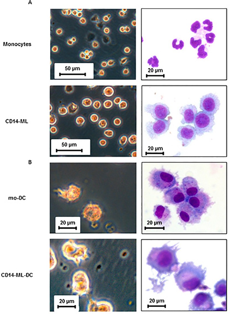
Fig 1. Morphology of CD14-ML and CD14-ML-DC generated by the current procedure.

(A) Phase-contrast images of live cells (left) and cytospin samples stained with May–Grünwald Giemsa (right) of the human monocytes and monocyte-derived myeloid cell lines (CD14-ML) are shown. (B) Morphology of OK432-stimulated mo-DC and CD14-ML-DC are shown. mo-DC and CD14-ML-DC were stimulated with OK432 for 2 days and subjected to microscopic analysis. The data are representative of 2 experiments.
