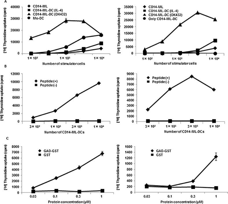
Fig 3. T-cell stimulation and antigen presentation by CD14-ML-DC.

(A) CD14-ML (rhombuses), CD14-ML-DC stimulated with IL-4 (squares), OK432 (triangles), or mo-DC (circles) were irradiated and co-cultured with allogeneic peripheral blood T cells (4×10⁴ cells/well) or alternatively, only CD14-ML-DC (circles) were irradiated and cultured in a 96-well round-bottomed culture plates for 5 days. T cell proliferation was measured according to [3H]-methylthymidine-uptake in the last 16 h. The experiments were conducted on CD14-ML derived from 2 different donors. (B) The indicated numbers of CD-14-ML-DC were loaded with GAD65111-131 peptide (rhombuses) or those left unloaded (squares), X-ray-irradiated, and co-cultured with GAD65-specific HLA-DR53-restricted clonal human CD4+ T cells (3×10⁴ cells/well) for 3 days. The proliferative response of the T cells was measured by incorporation of [3H]-methylthymidine in the last 16 h of culture. The experiments were conducted on CD14-ML derived from 2 different donors. (C) CD14-ML-DC were pulsed with recombinant GST-fused GAD65 protein or GST protein for 16 h, X-ray-irradiated, and subsequently added to GAD65-specific T cells. The proliferative response of the T cells was analyzed. The experiments were conducted on CD14-ML derived from 2 different donors.
