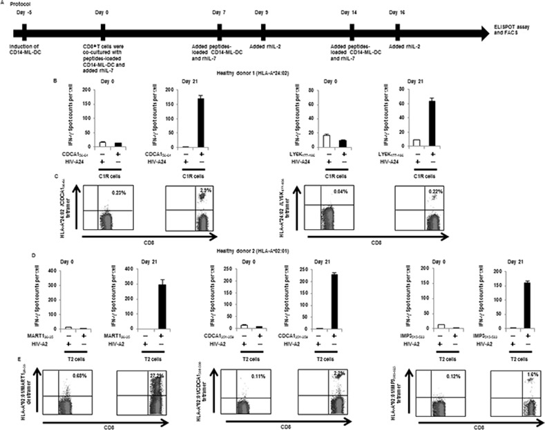
Fig 4. Induction of CD8+ T cell lines that are reactive to cancer antigens by CD14-ML-DC.

(A) Protocol for the induction of cancer antigen-specific CD8+ T cells by CD14-ML-DC. In order to generate CD14-ML-DC, we added IL-4 to CD14-ML. After 3 days, we added OK432. CD14-ML-DC were pulsed with peptides for 3 h, X-ray-irradiated (45 Gy), and subsequently mixed with autologous CD8+ T cells. Cells were cultured with rIL-7 (10 ng/ml) in AIM-V with 5% human decomplemented plasma. On days 7 and 14, the T cells were restimulated with the autologous peptide-pulsed CD14-ML-DC and on days 9 and 16, and were supplemented with rIL-2 (20 IU/ml). CD14-ML-DC were prepared each time, and we only added IL-4 (did not add OK432). IFN-γ ELISPOT assay and flow cytometry were performed after 6 or 7 days from the third round of peptide stimulation. (B, C) Peripheral blood CD8+ T cells were obtained from a HLA-A*24:02-positive healthy donor (healthy donor 1) and were co-cultured with 4 peptides (CDCA156-64, KIF20A66-75, LY6K177-186 and IMP-3508–516)-loaded autologous CD14-ML-DC. (B) On day 21, the number of IFN-γ producing CD8+ T cells were analyzed by ELISPOT assay (Day 21). The results of the T cells before stimulation culture are also shown (Day 0). The HIV-peptide was used as a control peptide. (C) On day 21, the T cells were recovered and stained with anti-CD8 mAb and the HLA-A*24:02/CDCA156-64 or HLA-A*24:02/LY6K177-186 tetramer. The numbers in the figure indicate the percentage of the CD8+ T cells that were positively stained with the tetramer of the HLA-peptide complex (Day 21). The results of the T cells before stimulation culture are also shown (day 0). (D, E) A similar experiment as in (B, C) was done with the cells obtained from a HLA-A*02:01-positive donor (healthy donor 2). We used 4 peptides (CDCA1351-359, KIF20A809-817, MART126-35 and IMP3515-523) for the stimulation of the T cells. (D) The number of IFN-γ producing CD8+ T cells was analyzed by ELISPOT assay. (E) The T cells were recovered and stained with an anti-CD8 mAb and a HLA-A*02:01/MART126-35 dextramer, HLA-A*02:01/CDCA1351-359 tetramer or HLA-A*02:01/IMP3515-523 tetramer. The numbers in the figure indicate the percentage of the CD8+ T cells that were positively stained with the dextramer or tetramer of HLA-peptide complex.
