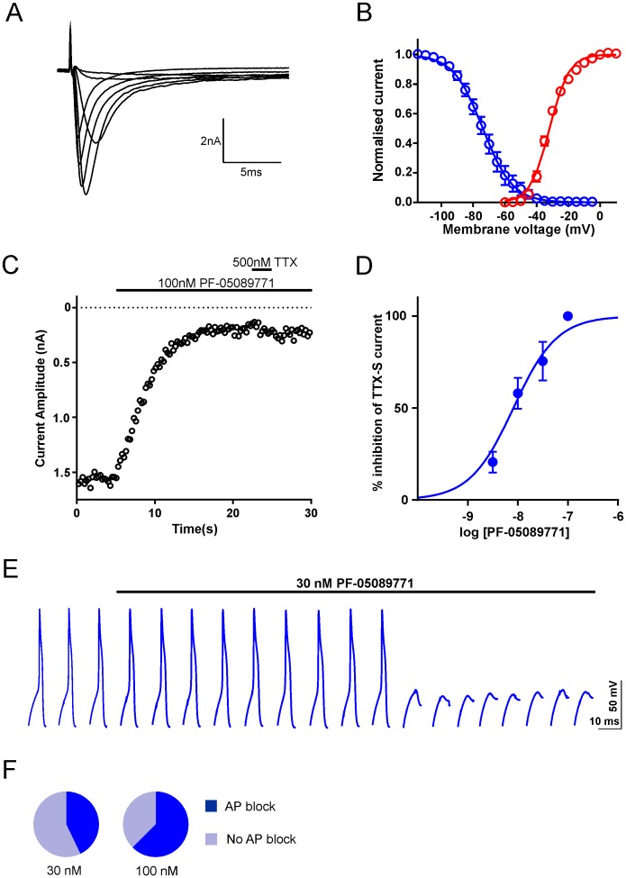
Fig 5. Evidence for functional Nav1.7 in human DRG neurons.
A. Representative TTX-S current traces (recorded in the presence of 1 μM A-803467 and following graded voltage steps from -110 mV to 10 mV. B. Voltage dependence of activation (red, n = 4 for each voltage) generated from the protocol described in A and steady state fast inactivation (blue) generated by conditioning 500 msec prepulses to voltages between -110 mV and +10 mV followed by a test pulse to 0 mV from a holding potential of -110 mV (n = 4 for each voltage). Both datasets are fitted with Boltzmann functions. C. Representative timecourse relationship for peak TTX-S current following the application of 100 nM PF-05089771 and 500 nM TTX. D. Concentration-response relationship for PF-05089771 block of TTX-S current (IC50, slope: 8.4 nM, 1.1; n = 3–6 per concentration) E. Example voltage traces from a current clamp recording. Single action potentials were evoked by a 20 ms suprathreshold current step at 0.1 Hz. The scale bar refers to the voltage traces whereas the start-to-start interval is 10 s. F. Summary pie charts showing that the application of 30 and 100 nM PF-05089771 resulted in action potential block in 3/7 and 5/8 DRG neurons respectively.

