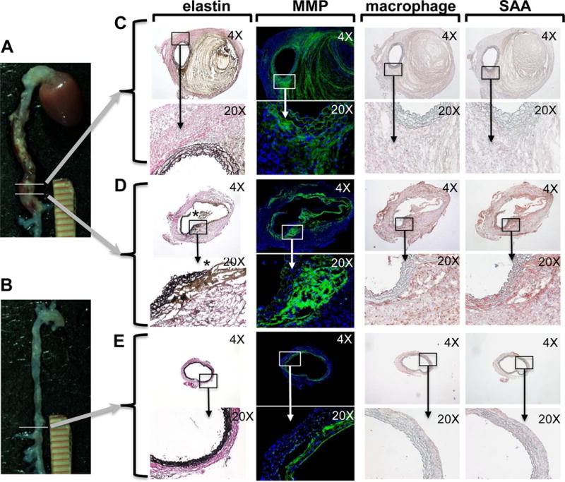
Fig 3. SAA is present in AngII-induced AAA and co-localizes with macrophages, elastin breaks and MMP activity.

Male apoE−/− mice were infused with 1,000 ng kg−1 min−1 AngII for 10 days. A) Mouse that developed AAA and B) non-responsive mouse. Adjacent sections from two different regions of AAA (denoted by horizontal lines) were analyzed in parallel to allow direct comparisons. C, D) Sections were processed to detect elastin fibers by a modification of Verhoeffs staining (* denotes region with elastin breaks); MMP activity by in situ zymography (green fluorescence); and macrophages and SAA by immunostaining (red chromogen), and then photographed under 4× and 20× objective magnification as indicated. For in situ zymography, nuclei were identified using DAPI (blue fluorescence). E) Sections from a non-responsive mouse processed to detect elastin fibers, MMP activity and immunostaining for macrophages and SAA.
