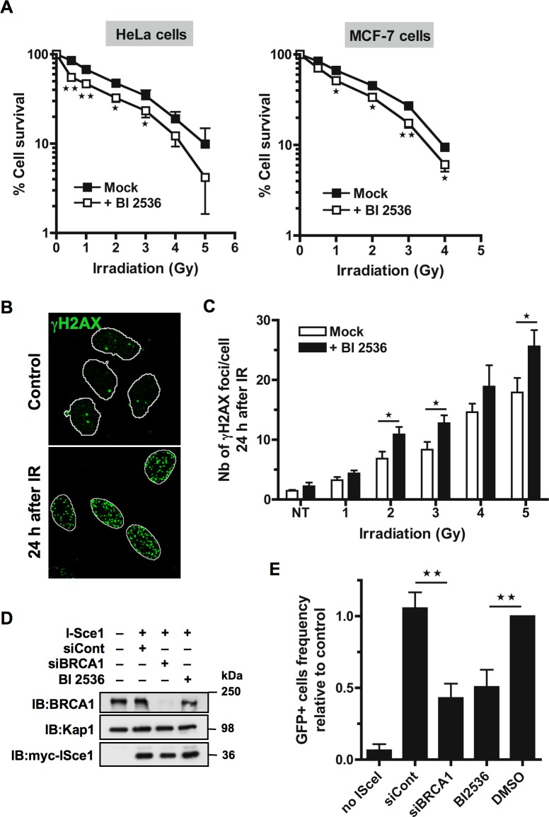
Figure 1. Inhibition of Plk1 sensitizes cells to ionizing radiations.
A. HeLa and MCF-7 cells are radio-sensitized when Plk1 is inhibited before IR. Cells were pre-treated or not with 10 nM BI2536 for 2 h before exposure to a range of doses of IR (0-5 Gy). Colonies were stained 10 to 12 days following IR and counted. Graph shows the mean percent surviving at each dose of IR relative to colonies formed at 0 Gy ± SE over 3 independent experiments done in triplicate. Significant differences in cell survival were assessed using a two-tailed paired Student's t-test and are indicated by * = p < 0.05; ** = p < 0.01.B. The incidence of γH2AX foci was determined in HeLa cells that were pre-treated or not with BI2536 for 2 h and then mock-exposed or exposed to IR, washed and collected 24 h after to perform immunofluorescence assay. Cells were immunostained with γH2AX antibody, probed with DAPI and then examined by confocal fluorescence microscopy. Representative images of γH2AX foci (green) in mock-exposed nuclei (control) or in irradiated nuclei 24 h following IR (4 Gy) are shown. C. The incidence of γH2AX foci is increased 24 h post-IR when Plk1 is inhibited. The number of foci was quantified using ImageJ software (NIH). Graph shows the mean number of γH2AX foci ± SE per cell over 3 independent experiments, n ≥ 100 cells per time-point. Significant differences in γH2AX foci number were assessed using a two-tailed unpaired Student's t-test and are indicated by * = p < 0.05. D. BRCA1 down-regulation by siRNA and expression of myc-tagged I-SceI endonuclease were analyzed after transfection with control siRNA (siCont) or siRNA against BRCA1 (siBRCA1) and subsequent transfection with myc-tagged I-SceI encoding plasmid (I-SceI) in presence or absence of Plk1 inhibitor (BI2536). Whole-cell extracts obtained from HEK293/DR-GFP cells were resolved by SDS-PAGE and immunoblotted using anti-BRCA1 and anti-myc antibodies. Equal loading was confirmed using anti-Kap1 antibody. E. DSB repair by HR is reduced in presence of Plk1 inhibitor. Frequencies of GFP-positive cells were measured by FACS following transient I-SceI expression for HEK293/DR-GFP cells treated with Plk1 inhibitor (BI2536) or mock-treated (DMSO), along with HEK293/DR-GFP cells transfected by siCont or siBRCA1. Graph shows the frequency of GFP-positive cells ± SE relative to the mean value of control DMSO-treated cells, n = 5. Significant differences in HR efficiency were assessed using a two-tailed paired Student's t-test and are indicated by ** = p < 0.01.

