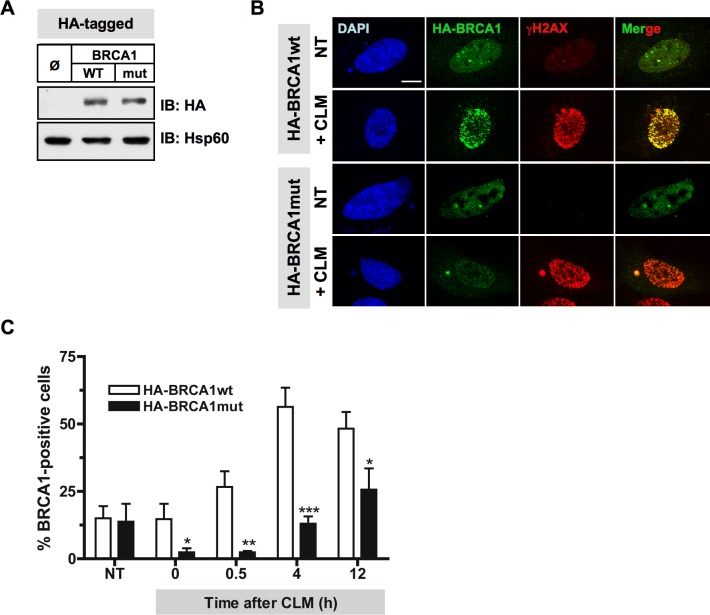
Figure 6. Mutations of Plk1 sites on BRCA1 compromise BRCA1 foci formation following DSB.
A. Immunoblotting using anti-HA and anti-Hsp60 antibodies indicates equivalent amounts of the HA-tagged BRCA1 proteins expressed in HeLa cells. B. BRCA1 foci formation induced by DNA damaging treatment is impaired when Plk1 sites are mutated. 24 h following transfection with HA-tagged BRCA1 constructs, HeLa cells were left untreated or treated with calicheamicin (CLM) for 1 h, washed and collected at different time-points following treatment to perform immunofluorescence assay. Cells were immunostained with anti-HA and anti-γH2AX antibodies, probed with DAPI and then examined by confocal fluorescence microscopy. Representative images of HA-BRCA1 (green) and γH2AX (red) co-staining in untreated cells (NT) or in CLM-treated cells 4 h after treatment are shown. C. The number of BRCA1-positive cells upon CLM treatment is reduced when BRCA1 is mutated at S1164/S1377. The number of foci was quantified using ImageJ software (NIH). Graph shows the mean number of positive cells containing more than 5 BRCA1 foci ± SE over 3 independent experiments, n ≥ 120 cells per time-point. Significant differences in BRCA1-positive cells numbers were assessed using a two-tailed unpaired Student's t-test and are indicated by * = p < 0.05, ** = p < 0.01 and *** = p < 0.001

