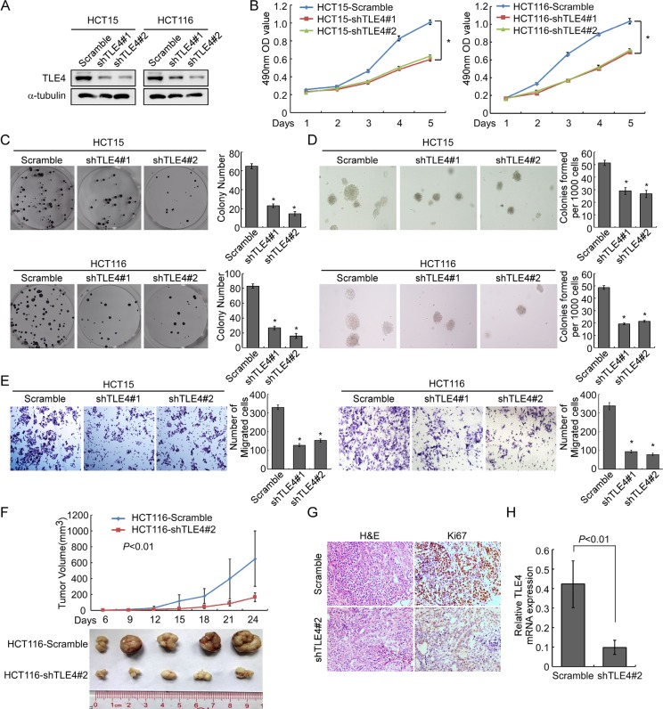
Figure 4. Depletion of TLE4 inhibits cell proliferation and invasion activity.
(A) RNAi-silencing of TLE4 in shRNA-transduced stable HCT15 and HCT116 cells. α-Tubulin was used as a loading control. (B and C) Reduction of endogenous TLE4 inhibited cell growth in MTT assays (B) and colony formation assays (C) *P < 0.01. (D) Silencing of TLE4 inhibited cell growth ability of HCT15 and HCT116 in Soft agar colony formation assays. Colonies containing more than 50 cells were scored. Error bar represents the mean ± SD of three independent experiments *P < 0.01. (E) The invasive abilities of CRC cells evaluated using the Matrigel-coated Boyden chamber invasion assay. Each bar represents the mean ± SD of three independent experiments. *P < 0.01. (F) Xenograft model was built by injected HCT116/vector and HCT116/shTLE4 cells in nude mice (n = 5/group). Tumor volumes were measured on the indicated days. Data points are the mean tumor volumes ± SD. (G) The sections of tumor were under H & E staining or subjected to IHC staining using an antibody against Ki-67. (H) Real-time PCR was used to test TLE4 expression in xenograft tumors formed from HCT116/Scramble and HCT116/shTLE4. Error bar represents the mean ± SD.

