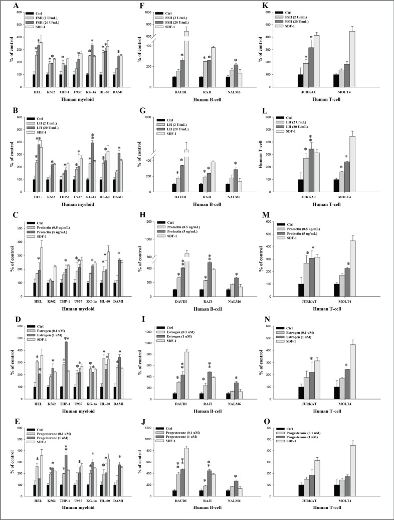
Figure 3. Pituitary and gonadal sex hormones enhance the adhesiveness of human leukemia cells to fibronectin.
(A–E) Adhesion of myeloid leukemia cell lines to fibronectin-coated surfaces in response to FSH (2–20 IU/mL), LH (2–20 IU/mL), prolactin (0.5–5 μg/mL), estradiol (0.1–1 μM), and progesterone (0.1–1 μM). A similar experiment was also performed to assess the effect of these hormones on the adhesion of different B-lymphoid (F–J) and T-lymphoid (K–O) leukemic lineages. Quiescent cells (3000 cells/100 μL) were stimulated with these hormones at the indicated concentrations in medium with 0.5% BSA for 5 min incubation at 37°C. After the non-adherent cells were removed via three consecutive washes, the number of adherent cells was measured by microscopic analysis. The effects of all these hormones on adhesion of all employed leukemia cell lines were also evaluated for adhesion versus stromal-derived factor 1 (300 ng/mL) and RPMI medium containing 0.5% BSA as a positive and negative control, respectively. The negative control values are normalized to 100%. Data are displayed as means ± SD, with a statistical significance *p ≤ .05 and **p ≤ .01 versus unstimulated control cells.

