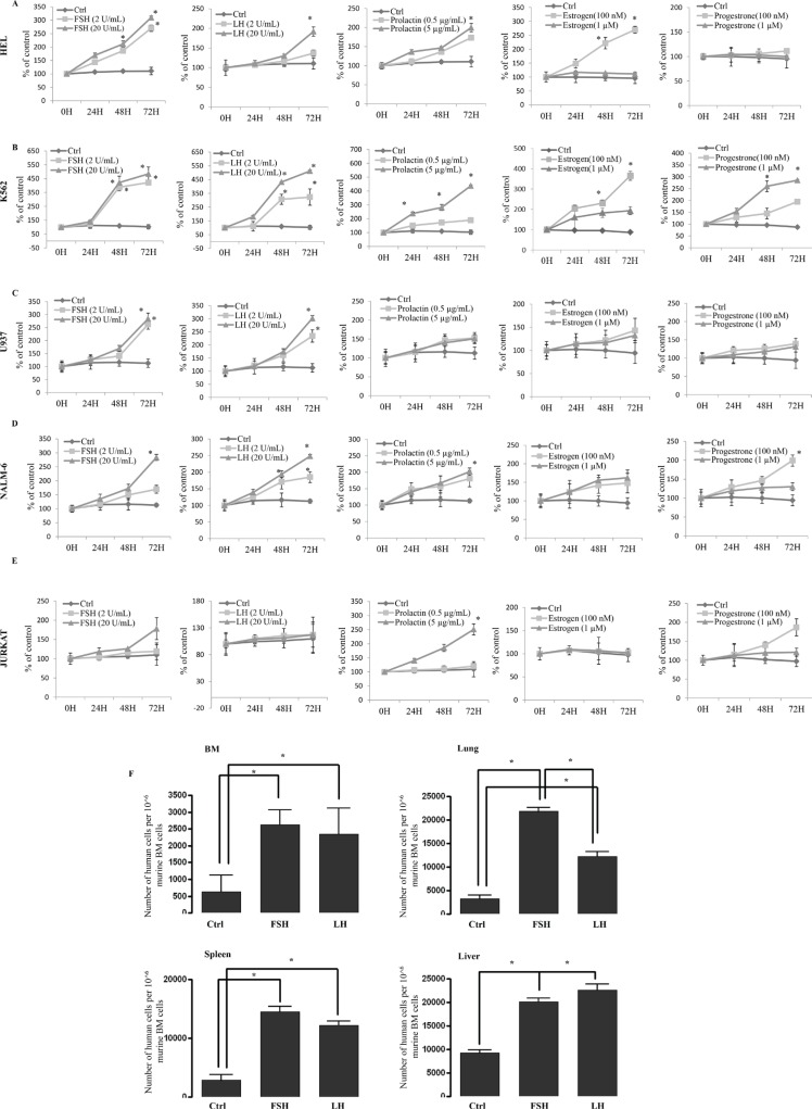
Figure 4. Human myeloid and lymphoid leukemia cells proliferate in vitro and migrate in vivo in response to SexHs.
(A–C) Proliferation of human myeloid (erythroblastic [HEL, A], erythrocytic [K562, B] and monoblastic/cytic [U937, C] leukemia cells stimulated by pituitary and gonadal SexHs in a dose-dependent manner. (D–E) Proliferation of human B-lymphoid (lymphoblastic [NALM-6, D]) and T-lymphoid (lymphoblastic [JURKAT, E]) leukemia cells by pituitary and gonadal SexHs, in a dose-dependent manner. All proliferation experiments were done in RPMI culture medium containing 0.5% BSA for 72 hours using 0.3 × 10⁴ cells/well in a 96-well plate. The negative control values are normalized to 100%. For each cell line, the experiment was repeated twice in triplicate with similar results. (F) Detection of in vivo-transplanted FSH- or LH-treated human myeloid THP-1 leukemic cells in the organs of irradiated mice post transplantation. As shown, the number of FSH- or LH-treated leukemic cells was significantly higher in isolated organs from mice than in ex vivo-untreated leukemic cells. Detection was performed by employing RT–qPCR for the presence of human Alu sequences in purified genomic DNA samples. For statistical comparisons, a one-way analysis of variance and a Tukey's test for post hoc analysis was carried out, and means ± SD are shown. Significance levels, *p ≤ .05 versus control.

