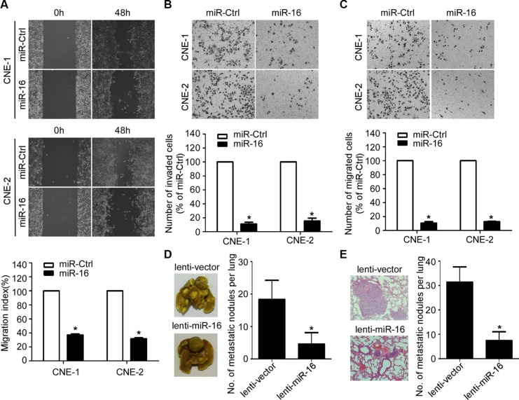
Figure 3. miR-16 inhibits NPC cell migration, invasion, and lung metastatic colonization.
(A) Representative images and quantification of wound healing assays in the indicated cells. (B–C) Representative images and quantification of the indicated cells determined by Transwell migration (B) and invasion assays (C). (D–E) Lung metastatic colonization models in nude mice were constructed. Representative images and quantification of macroscopic tumor nodules formed on the lung surface (D); as well as of microscopic tumor nodules formed in the lung tissue sections stained with hematoxylin and eosin (E) are shown. The data are presented as the mean ± S.D.; p values were calculated using Student's t-test.

