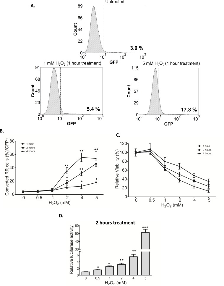
Figure 2. RU cells converted to RR cells upon H2O2 challenge.
A. RU cells derived from MCF7 were exposed to varying doses of H2O2 for 1 hour in serum free media. Flow cytometry was used to assess the expression of GFP in the viable cell populations. Data is expressed relative to untreated negative control cells and the values represent the GFP positive cells. Addition of H2O2 to RU cells increased the proportion of GFP-positive cells (from 3.0%, background level to 17.3%). B. Data is expressed as percent of cells with higher GFP expression relative to untreated negative control detected by flow cytometry (called converted RR cells/GFP+) after exposure to varying doses of H2O2 for different time points in serum free media. The proportions of converted RR cells (or GFP-positive) significantly increased in a time- and dose-dependent fashion. C. Cells from above experiments were subjected to MTS assay to assess the cell viability at the end of experiments. Data is expressed as percentages of the negative control cells, which were set as 100%. The cell viability decreased in a time- and dose-dependent fashion. D. RU cells derived from MCF7 were exposed to varying doses of H2O2 for 2 hour in serum free media. Data is expressed as luciferase activity relative to untreated negative control. RU cells treated with H2O2 significantly increased the luciferase activity in a dose-dependent manner.

